A NEW KIND OF ICE AGE
By: Allessandra DiCorato
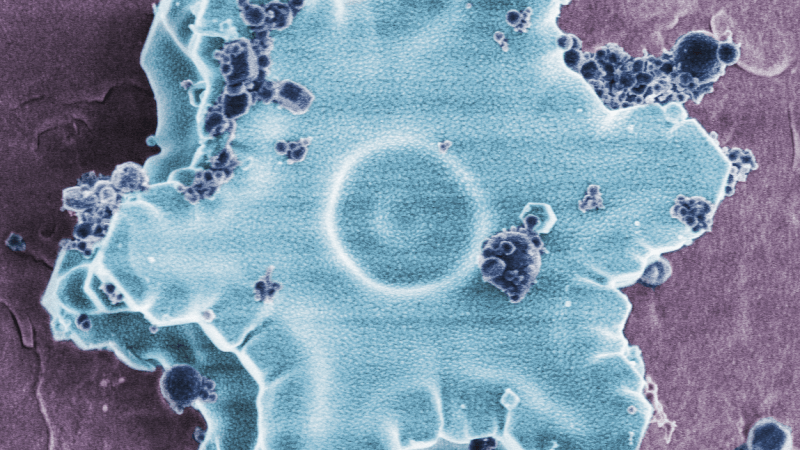
False colored ice. Image is courtesy of Allessandra Dicorato. Editing by Ridvan Kahraman
Using cryoEM, amorphous ice allows scientists to observe specimens in a near-natural state
Ice often evokes images of cold drinks on a summer’s day, icicles dripping off a roof in winter, or frost spreading on a windowpane. Its appearance and texture, most will agree, are familiar: the slippery layer beneath a car wheel as you drive home late on Christmas eve, or the pond you skated on as a child.
In reality, the ice you and I think of in the natural world is just one of many types. This familiar kind of ice forms when water molecules align in ordered, hexagonally packed crystals. Under a high-powered microscope, these crystals resemble a mixture of what you’d expect — micrometer-sized snowballs or six-fingered stars — but also exotic cuboidal crystals and ferns. These shapes are of the same crystalline structure, or phase, as frozen ponds.
By actively imposing a different structure — amorphous ice — in their samples and maintaining those specimens at around -200°F, scientists are using freezing in a whole new way to preserve biology in a pristine state. These methodologies allow one to probe the smallest biological processes in research that is beautiful in its precision, the patience it demands, and the detail it affords.
Amorphous ice is far less common than ice’s crystalline forms, but far more useful to the electron microscopist. In the natural world, this phase exists in upper atmosphere clouds only visible at twilight, and on the surfaces of distant icy moons. While a crystal consists of atoms aligned like bricks, the molecules in amorphous ice are arranged randomly, leading to a solid phase that resembles glass and lacks snow’s granular shapes. This structure forms when water is subjected to extreme pressures or temperatures in the laboratory or during celestial events, and can be used to preserve a biological sample in what is as close to its native state as modern scientists can achieve. As simple as it may sound, the discovery of this preservation method in the 1980’s earned Jacques Dubochet the 2017 Nobel Prize in Chemistry.
Just as one must proceed cautiously on an icy road, even with years of driving experience and a well-equipped car, so too must microscopists navigate the complex sample preparation required for preserving amorphous ice. Biological samples, such as human cells, tissue, or even an entire fruit fly, contain about 70 percent water. This renders them troublesome to prepare for an electron microscope, where a beam of electrons damages specimens. In the past, scientists used chemical fixatives to preserve nanoscale details as small as the cell membrane, 10,000 times thinner than the width of a human hair, but even these techniques cause damage. Cryo-electron microscopy (cryoEM), in which biological samples are converted into amorphous ice, is often a biologist’s only hope at observing an unaltered moment in time.
With the sound of a gunshot and a kind of mechanically shuddering sigh, high-pressure freezers spit out samples frozen in amorphous ice in seconds. Only once the sample is frozen does the truly precise work begin. Electron microscopes are maintained at sub-zero temperatures, so each piece of equipment that touches the specimen, from tweezers to storage containers, must be pre-cooled to avoid warming and refreezing the sample in damaging crystalline ice. If you breathe on your sample, or if it is particularly humid, at the microscope, you will find a lacy ice coating obscuring the nanometer-sized details you so painstakingly preserved three hours earlier. Samples themselves are only 3 millimeters in diameter and a fraction of that deep; they are constantly submerged in liquid nitrogen, which boils at room temperature and can shift a sample if improperly secured.
The care this low temperature environment demands means that preparing a sample for observation can take hours, or, depending on a chosen method’s nuances, even days, and adds cost to an already expensive experiment. (A process known as freeze substitution, for instance, can involve dehydrating a sample for four days and infiltrating its newly porous depths with liquid resin, which can take up to six days to stiffen.) Furthermore, contrast in an electron microscope depends upon differences in composition. Because biological samples are mostly water, without staining cellular features, an electron microscopist is left to stare bleakly at a largely gray screen, hoping for a discernable feature. Finally, once a single imaging session is complete, it can produce so much data that it is difficult to process with most software.
And yet, there is a kind of poetic stillness in observing a cell caught mid-division with each structure frozen in place, its innermost mechanisms suspended in time. It is the closest that we come to seeking answers from biological creatures too small to study directly, and the only chance of glimpsing processes that occur in what seems to us an instant. This is all to say nothing of the ability to visualize single molecules where previous generations of scientists squinted at a blur. What cryoEM may teach us more than anything else, however, is the value of a single, exquisitely executed experiment. With no room for heavy breathing, warm tweezers, or shaky hands, cryoEM demands a biologist’s best, and elevates the deceptively simple task of observation to a frozen art.
Allessandra DiCorato is a rising fourth year PhD student in the Materials Science Department, where she works in the Joester lab to develop cryo-imaging techniques to study the interactions between nanomaterials and cancer cells. After graduating, she hopes to pursue a career that combines her two interests, science and writing.